Celebrating Black History Month: Medicinal Practices of Enslaved Peoples

By Phoebe Warren, Center for Education Intern

The Center for Education celebrated Black History Month and the 2022 theme of health and wellness through a series of Instagram posts honoring the herbal medicinal traditions of enslaved African and African American healers. We’re publishing the content of the entire series here. Many of the plants we feature can be found during the warmer months in the Benjamin Rush Medicinal Plant Garden here at the College. Please visit us if you’d like to see these plants in person!

These posts touch on several themes including respiratory illness, spiritual empowerment, the sources of enslaved people’s medicinal knowledge, and reproductive health. All these themes demonstrate the resilience and wisdom of Africans and African Americans who used the medicinal powers of plants to do what they could to support their health and wellbeing in the oppressive, dangerous, and dehumanizing realities of slavery.

If you are interested in learning more about African American healers and plant-based remedies, Herbert Covey’s book African American Slave Medicine: Herbal and Non-Herbal Treatments (2007) is an excellent starting point. The Federal Writers’ Project Slave Narratives, available online through the Library of Congress, is one of the most extensive series of interviews of formerly enslaved African Americans. These interviews were conducted by writers in the late 1930s with funding from the Works Progress Administration. The narratives are complicated historical sources. For example, there was often a cultural and racial disconnect between the writers and the people interviewed. Nevertheless, the project is one of the most extensive efforts to document the experiences and memories of formerly enslaved people.

Disclaimer: It’s important to note that some of the plants featured below are toxic and are not recommended for use. Please consult a medical professional before using any herbal supplement.

Image Credit: Karen E. Price, Mount Vernon Preservation

Knowledge of the curative powers of plants was a way for Africans and African Americans to survive and care for themselves and their loved ones. Plant knowledge and ritual practices around health and healing were passed down through generations.

This vessel, found on the Curriboo Plantation in South Carolina, was made by hand by enslaved people and was used for preparing food and medicines. Similar handmade pottery has been found throughout the American South. Some are marked with symbols similar to cosmograms, sacred symbols found in West and Central African spiritual traditions.

Image credit: Dionysia CC BY-SA 3.0

Horehound – Marrubium Vulgare

Respiratory diseases like pulmonary tuberculosis, whooping cough, and bronchitis affected the health of enslaved Africans and African Americans. In Federal Writers’ Project interviews in the 1930s, formerly enslaved people remembered using horehound to soothe sore throats and coughs. Sally Murphy of Alabama recalled, “When any of us got sick, we was give horehound tea and rock candy.” Horehound is native to North Africa, Europe, and Central Asia, and is part of the mint family. Horehound contains chemical compounds that act as expectorants to loosen and clear mucus from the airway.

Image Credit: Agnieszka Kwiecień, CC BY-SA 4.0

Ferula species

Ferula species (possibly Ferula foetida), which formerly enslaved people referred to as “asafetida,” were also used to treat respiratory illness. These plants are members of the carrot family. Today, the term asafetida usually refers to dried latex prepared from the plant’s roots. Ferula species have a strong odor and are also known as “stinking gum” or “Devil’s dung.” Africans and African Americans used asafetida as a preventative against respiratory diseases. It was thought that breathing in the plant’s odors would kill disease-causing material. Lizzie Norfleet described her experiences under slavery in an interview with the Federal Writers’ Project: “Every child wore an asafetida bag round the neck to keep from ketching diseases.”

Elderberry or Elder · Sambucus sp. (Sambucus canadensis, Sambucus nigra)

Many Africans and African Americans believed in the power of time-honored remedies and trusted healers to cure illnesses. By growing and foraging for medicinal herbs, enslaved people were empowered to heal themselves and their loved ones. However, if those they cared for did not recover, enslaved healers were often blamed by white overseers and enslavers. Laws enacted by the Virginia legislature as early as 1748 determined that enslaved people administering medicines without approval by their enslaver was an act punishable by death. Sacred practices for health and healing, sometimes conducted in secret, were a means of survival for Africans and African Americans enduring the cruelties of enslavement.

In Federal Writers’ Project interviews, formerly enslaved African Americans recalled using elderberry, also called elder, as a powerful preventative and remedy. Harriet Collins of Texas remembered that elder was placed around infant’s necks to prevent discomfort when teething. Rachel Goings of Missouri described a remedy for sores: “I’d a took elder leaves en boiled em to make a tea—den I’d poured dat in de sore and it ud got well.”

Coneflower or Sampson root · Echinacea sp.

Enslaved Africans and African Americans learned about local medicinal plants from Indigenous healers. Native Americans had and continue to have an intimate knowledge of local plants. Native Americans were sometimes enslaved alongside Africans, and some Native American towns sheltered people seeking freedom from slavery. Knowledge about medicinal plants was likely shared in these contexts.

Coneflower is native to North America. Native Americans used the plant to treat gastrointestinal issues, fevers, sore throats, toothaches, and burns. In interviews with the Federal Writers’ Project, formerly enslaved African Americans called this plant Sampson root. Phil Town of Georgia remembered using a Sampson root tea to cure cramps. Pierce Harper of Texas and Fannie Moore of North Carolina both used the plant to treat stomach pain.

Okra · Abelmoschus esculentus

African people forced into enslavement had a rich knowledge of the medicinal powers of plants. The indigenous plants of the Caribbean and American South were very different from those of West and Central Africa. But some African plant species were brought to the Americas as a result of the transatlantic slave trade. Some of the captured African people who survived this unimaginable journey wore strings of plant seeds, such as wild licorice. Enslavers lined the holds of slave ships with African grasses. Crops brought from Africa like okra, yams, and black-eyed peas were staple foods for enslaved Africans and African Americans.

Okra was also used for medicinal purposes. Katie Arbery, a formerly enslaved African American who was interviewed through the Federal Writers’ Project, described the healing power of okra. Arbery remembered a time when she was so sick that, “They give me up to die once.” After trying different medicines with no success, her doctor “fed me for three weeks steady on okra soup cooked with chicken…Then I commenced gettin’ better and here I am.”

Image credit: Wellcome Collection, CC BY 4.0. This image is from a book by 19th-century British botanist Mary Ann Burnett titled Illustrations of useful plants employed in the Arts and Medicine. You can find it in our Historical Medical Library

Cotton root · Gossypium herbaceum

Many enslaved African and African American women were sources of medicinal knowledge, and many served as midwives and healers. Using plant-based remedies and knowledge gained from experience, enslaved midwives delivered babies and did what they could to alleviate complications with pregnancy and childbirth. Enslaved women used different plants as contraceptives, abortifacients, and to regulate menstruation, induce labor, and ease labor pains.

Enslaved women’s reproductive health was threatened by inhumane conditions, malnutrition, rape, and violence. The plantation system pressured enslaved women to have as many children as possible. In interviews with the Federal Writers’ Project, formerly enslaved African Americans described that in secret, enslaved women used the root of the cotton plant to prevent pregnancy. When 19th-century white physicians and enslavers became aware of this practice, it was seen as a threat. Although enslaved women were denied the right to have control over their reproductive lives, they resisted by using plant-based medicines and other methods to assert agency over whether and when they had children.

Sage · Salvia officinalis

The medicinal practices of enslaved Africans and African Americans have been systematically left out of the historical record. In the 18th and 19th centuries, white legislatures passed laws limiting enslaved people’s access to plants and preventing them from practicing medicine. Enslaved people were often prevented from learning how to read or write. The institution of slavery separated families from one another and purposefully disrupted the passing down of generational knowledge.

Today, thanks to the work of researchers, community leaders, and libraries, these histories are being told. Please look out for and share these stories, and support Black healers, botanists, and gardeners in your community! It’s our hope that this series will encourage us to look differently at the plants we encounter, including herbs like sage. Sage was used by enslaved people as a cure for fever, chills, or, as Mrs. Mary Kincheon Edwards of Texas remembered, “Some people would use it fo’ almost anything.”

Bibliography

Aćimović, Milica et al. “Marrubium vulgare L.: A Phytochemical and Pharmacological Overview.” Molecules 25, no. 12. June 24, 2020.
Andreae, Christine. “Slave Medicine.” Jefferson Library at Monticello.
Covey, Herbert C. African American Slave Medicine: Herbal and Non-Herbal Treatments. Lexington Books, 2007.
Eisnach, Dwight, and Herbert C. Covey. “Slave Gardens in the Antebellum South: The Resolve of a Tormented People.” The Southern Quarterly 57, no. 1 (2019): 11-23.
Federal Writers’ Project: Slave Narrative Project, Vol. 2, Arkansas, Part 1, Abbott-Byrd. November-December, 1936.
Ferguson, Leland & Kelly Goldberg (2019) “From the Earth: Spirituality, Medicine Vessels, and Consecrated Bowls as Responses to Slavery in the South Carolina Lowcountry.” Journal of African Diaspora Archaeology and Heritage, 8:3 (173-201). DOI: 10.1080/21619441.2019.1690843
Fett, Sharla. Working Cures: Healing, Health, and Power on Southern Slave Plantations. Chapel Hill: University of North Carolina Press, 2002. Pg. 63-65.
Native American Ethnobotany: A database of plants used as drugs, foods, dyes, fibers, and more, by Native Peoples of North America.
Owens, Deirdre Cooper. Medical Bondage: Race, Gender, and the Origins of American Gynecology, University of Georgia Press, 2017. Pg. 42-72.

Medicinal Orchids at the Mütter Museum

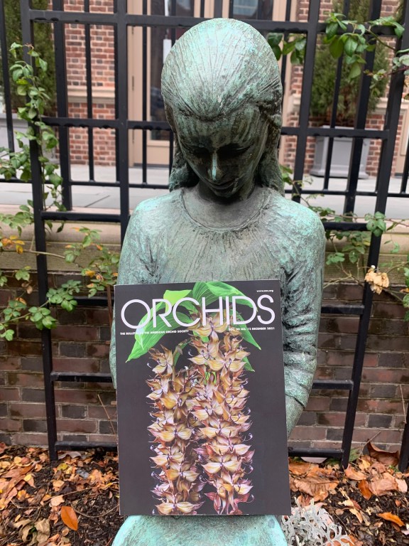
Hello, MütterEDU readers, my name is Jacqui Bowman, and I am the Co-Director of Living Exhibits at The Mütter Museum of The College of Physicians of Philadelphia. In the December issue of Orchids Magazine, it was announced that the Mütter Museum has received an education grant from the American Orchid Society to “create intrigue and educate visitors” about the genus Dendrobium and its historical and contemporary importance in medicine. This is the first of a series of articles that will share information about this project.

Bronze statue of a girl standing with bowed head. In the foreground positioned above her hands is a copy of Orchids magazine showing the front cover, depicting two orchids.

The Mütter Museum gets thousands of visitors of all ages every year (over 180,000 in 2019) and
many of them visit the Benjamin Rush Medicinal Plant Garden. Very few of them know anything about how orchids are used in medicine, or even about orchids at all – beyond the Phalaenopsis they see for sale at the supermarket down the street. Having orchids on display provides a tremendous opportunity to teach visitors about the extraordinary variety of orchids. It is also an opportunity to engage with communities in Philadelphia who may use orchid plants as medicine or recall their families doing so.

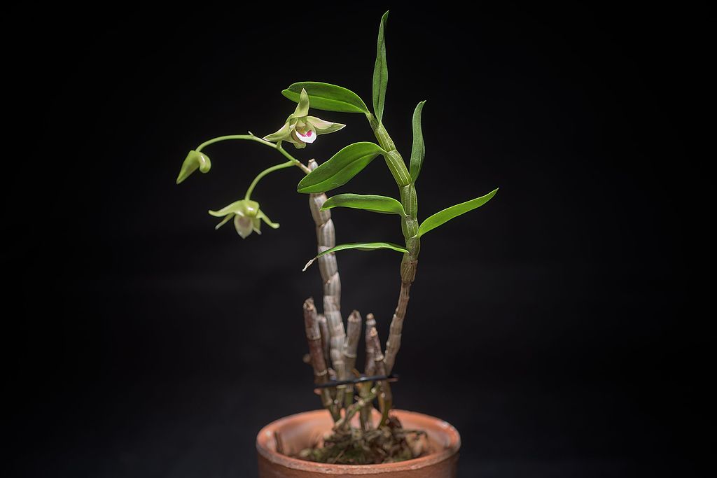
Photograph of a Dendrobium catenatum.
Dendrobium catenatum (Image credit: snotch via Wikimedia Commons; used under CC BY 2.0; no changes made)

The genus Dendrobium is one of the largest orchid genera and has enormous economic
importance globally as a source of ornamental plants as well as medicine. These orchids are
used widely in India and Asia and by communities all over the world who trace their roots to
these geographic areas. The use of orchids in medicine has the longest history in China where
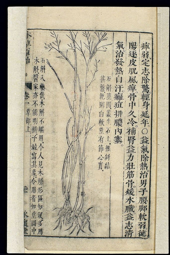
Dendrobium moniliforme (L.) sw. and Dendrobium catenatum Lindl. have been used for at least 1000 years. Dendrobium (Shihu) was described in the first Chinese Materia Medica, Shen Nong Bencao Jing, in the first century C.E. They are an enormously variable genus in terms of size and appearance. At least 70 of them have been investigated for pharmacological properties that have been shown to impact disease processes including cancer and diabetes.

Photograph of a bag from the Chinese Materia Medica Shen Long Bencao Jing, with a drawing of orchid plants surrounded on both sides by Chinese characters.
Page from a Chinese Materia Medica depicting Dendrobium (Image Source: Wellcome Images via Wikimedia Commons; used under CC BY 4.0; no changes made)

The reason I initially became interested in Dendrobium orchids was through an interest in Japanese orchids known as Chouseiran or “long life orchids.” These are Dendrobium moniliforme (L.) orchids collected for their beauty and variety in shape and color of their leaves, canes, and flowers. They have been collected in Japan since the Edo Period (1603 – 1867), and they are typically displayed in beautiful ceramic pots. It was in reading about these orchids that I came to discover about how they, and many other Dendrobiums, have been used in medicine for thousands of years.

Last summer we were lucky to be able to borrow some beautiful Dendrobium orchids from The Barnes Arboretum of St Joseph’s University and display them in the Benjamin Rush Medicinal Plant Garden at the Mütter Museum. We are currently working on a small indoor cold-weather exhibit of some of the medicinal Dendrobium orchids for the interior of the building. As this project continues, we will share information via this blog and on Instagram at @MutterEdu #mutterorchids.

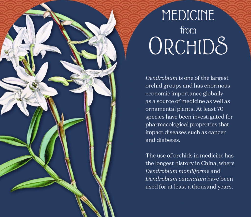
Museum exhibit label titled "Medicine from Orchids." On the left is an image of an orchid. On the right is the following text: "Dendrobium is one of the largest orchid groups and has enormous economic importance globally as a source of medicine as well as ornamental plants. At least 70 species have been investigated for pharmacological properties that impact diseases such as cancer and diabetes. The use of orchids in medicine has the longest history in China, where Dendrobium moniliforme and Dendrobium catenatum have been used for at least a thousand years."

Bibliography:

WFO (2021): Dendrobium catenatum Lindl. Accessed on 29 Dec 2021.

WFO (2021): Dendrobium catenatum Lindl. Accessed on 29 Dec 2021.

Cakova V, F Bonte, A Lobstein (2017). Dendrobium: Sources of Active Ingredients to Treat Age-Related Pathologies. Aging & Disease Vol 8 (827-849. December.

Medicinal Orchids of Asia by Eng Soon Teoh (2016) Springer International Publishing Switzerland.

Healing Orchids by Hong Hai & Soh Shan Bin (2020). World Scientific Publishing Co.Pte. Ltd.

The Relationship Between Art and Medicine

 Portrait of Samuel D. Gross. Source: Mütter Museum at The College of Physicians of Philadelphia Collections. 

Greeting, loyal MütterEDU readers, this fascinating article comes from Mütter Museum docent Angelie Cesario.

Within the Mütter Museum’s collection, there’s a portrait not often seen of Samuel David Gross. An American surgeon and teacher of medicine, Dr. Gross is best known for translating medical works into English and for his own publication on diseases of the bones and joints. At the Philadelphia Museum of Art, there’s a painting called The Gross Clinic by Thomas Eakins that depicts Gross standing in Jefferson Medical College’s surgical amphitheater demonstrating to a crowd his new procedure to treat bone infections. To his left, there are multiple doctors performing the surgery on the thigh of a patient while others in the crowd observe. At the time of its creation, the painting shocked and repelled viewers as many were not used to seeing such realistic art depicting gruesome surgery. Today, Eakins’ painting is still shocking but also appreciated by viewers. According to an art review in the New York Times, Eakins “painted, hands down, the finest 19th-century American painting.” (Kimmelman, 2002) 

 Thomas Eakins, The Gross Clinic, 1875, oil on canvas (Philadelphia Museum of Art & the Pennsylvania Academy of Fine Arts) 

The Gross Clinic raises important questions about the relationship between art and medicine. What other artists represented medicine and surgery in their art? Why were these representations important during the time in which they were created? Why are medical art and illustrations important today?   

One of the most renowned artists to have an impact on science and medicine was Leonardo da Vinci, a 16th century Italian artist, scientist, and engineer. Although probably best known for pieces such as The Mona Lisa and The Last Supper, he also specialized in many anatomical drawings. Performing numerous dissections during his time, da Vinci observed and studied human anatomy and physiology. He focused on the role of individual parts, including bones, muscles, and internal organs, as well as the mechanics behind them. Da Vinci recorded his findings during these studies through anatomical drawings which later contributed greatly to the advancements of both science and medicine. For example, at the Boston University School of Medicine, da Vinci’s anatomical drawings are still used in the classroom, providing students with accurate drawings to assist their surgeries.  

Human fetus, pen-and-ink studies by Leonardo da Vinci, c. 1510

Jacques-Fabien Gautier D’Agoty, an 18th century French anatomist and painter, was another influential artist during his time. In many of his paintings, he depicted realistic anatomical dissections of the human body. Some of Gautier D’Agoty’s paintings included up-close genitalia, a woman with an open uterus and fetus in her lap, and diseased individuals. Despite D’Agoty’s meticulous detail, experts in the medical field did not view his work as useful or educational. His work, instead, received greater attention for its style, which shocked contemporary observers. Regardless, D’Agoty still contributed greatly to the scientific world as an anatomist and philosopher through his scientific journals and publications.

 Jacques-Fabien Gautier D’Agoty, Female anatomical figure, Wellcome Library, London, UK. 

Jan Josef Horemans the Elder, an 18th century Flemish painter, is best known for his genre scenes of the everyday life of commoners. These paintings often depicted everyday activities, such as groups of people having discussions, listening to music, or playing cards. One painting, titled Interior with a Surgeon and his Apprentice Attending to a Patient, depicted a surgeon and his apprentice attending to a patient while observers looked in shock. In other paintings, Horemans the Elder depicts doctors attending to the sick, dentists extracting teeth extractions by dentists, and physicians performing other surgical procedures.  

Johan Joseph Horemans, Interior with a surgeon attending to a wound in a man’s side, c. 1722, Wellcome Collection, London, UK. (CC BY-NC 4.0)

When we look at the work of these artists, we can begin to understand that art reflects the changing scientific and cultural understandings of medicine. The Gross Clinic showed people learning in a group setting during a time where surgeries were often showcased to help people learn. Leonardo da Vinci, through dissections and his anatomical drawings and explanations, contributed greatly to the medical field years after his death. Jacques-Fabien Gautier D’Agoty showed how anatomical art moved from education to a more stylized, visual art. Jan Josef Horemans the Elder showed how medicine and surgery were practiced in everyday life of commoners.  

This brings us back to the question of why, in an era of modern medical technology, is medical-related art still important today? Today, medical art and illustrations work well for presenting multiple perspectives, allowing for easier learning through different colors and styles, and for dividing something whole into more understandable parts.       

One interesting recent example involves the study of COVID-19. In mid-January 2020, two medical illustrators at the Centers for Disease Control and Prevention were assigned the task of creating the first illustration of the SARS-CoV-2 virus, the virus responsible for the COVID-19 pandemic. Illustrations of the COVID-19 virus are important because they become a visual aid for a virus that’s essentially invisible to the human eye.  Since the first COVID-19 illustration publication, many more illustrations have been made that allow us to not only see what the virus looks like but also see the effects it has on different organs, how it can spread throughout the body, and how it can progress from moderate to severe cases. 

By creating illustrations, new medical discoveries can become more comprehensible to the general public and provide people outside of the medical field with a compelling visual aid. Through this we can see that medical art and illustration is still an essential part to education and learning, just as it was centuries ago. 

Sources: 

Bulletin of the Royal College of Surgeons. (2019, May 13). Jacques-Fabien Gautier D’Agoty – Exposition Anatomique des Organes des Sens, 1775. Royal College of Surgeons.  

Delbert, C. (2020, April 2). How illustrators created the iconic Coronavirus Image. Popular Mechanics.  

Encyclopædia Britannica, inc. (n.d.). Anatomical Studies and drawings of Leonardo da Vinci. Encyclopædia Britannica.  

Jargalsaikhan, B. B. (2021, July 16). Medicine in art: Wellcome library in London: DailyArt Magazine. DailyArtMagazine.com – Art History Stories.  

Kimmelman, M. (2002, June 21). A Fire Stoking Realism. The New York Times.  

May, T. (2020, August 20). 16 medical illustrators doing groundbreaking work. Creative Boom.  

Portrait of Dr. Samuel D. Gross (the Gross Clinic). Philadelphia Museum of Art. (n.d.).  

Riley, S. (2019, November 12). Anatomy professor uses 500-year-Old Da Vinci Drawings to guide cadaver dissection. PBS.  

Samuel David Gross. Biography. (n.d.). Retrieved October 13, 2021. 

The Junior Fellows Study Banned Books in the Historical Medical Library

The George and Judy Wohlreich Junior Fellows program is a three-year summer and after-school program for Philadelphia high school students from historically-excluded communities to learn about the diverse careers available in the healthcare and medical fields. We also feel it’s important for the healthcare professionals of the future to be active citizens; thus, we also include lessons that help promote their “civic health,” addressing such topics as voting rights, the social determinants of health, and systemic racism. To that end, we recently hosted a session devoted to Banned Books Week, a session that also gave us an opportunity to introduce the Junior Fellows to the Historical Medical Library.

Founded in 1788, the Historical Medical Library of The College of Physicians of Philadelphia was at one point Philadelphia’s central medical library, serving the city’s various hospitals and medical schools. Today, the Library is an independent research library devoted to the history of medicine and the medical humanities. The Library’s impressive collection includes over 400 incunabula (books published before 1501), historical archives and photographs, and over 12,000 books and rare texts related to the history of medicine (the Library also has a wonderful collection of digitized materials from the collection that are available to the public through the Digital Image Library).

Established by the American Library Association (ALA) in 1982, Banned Books Week celebrates the freedom to read by raising awareness of books that have been challenged or banned. While the First Amendment protects against the government banning books, individuals or organizations may attempt to limit access to certain forms of expression. One common method is to remove certain books from libraries, school reading lists, and curriculum. According to the First Amendment Encyclopedia, book banning is the most common form of censorship in the United States, and according to the ALA, 273 different books were challenged or banned in 2020 alone. The organization’s annual list of the top ten most challenged or banned books from the previous year included To Kill a Mockingbird by Harper Lee (for racial slurs and its depiction of the Black experience), George by Alex Gino (for discussions of LGBTQIA content), and Something Happened in Our Town: A Child’s Story about Racial Injustice by Marianne Celano, Marietta Collins and Ann Hazzard (for “divisive language” and allegedly promoting anti-police views).

During a recent session, the Junior Fellows met with members of The Historical Medical Library’s team to learn about the importance of libraries as repositories of knowledge, public servants, and forums for expression. Heidi Nance, Mary Hanes, and Kristen Pinkerton, the Library’s director, collections management librarian, and assistant librarian, respectively, shared their insights on a variety of subjects, including patron privacy, censorship, and cat paw prints on medieval manuscripts.

The Library staff also curated a selection of banned or challenged books from the collection. Among the more thought-provoking or surprising materials the Junior Fellows saw were Malleus Maleficarum, a fifteenth century guide to identifying and battling witches (for more, see our past article on the book) and The Immortal Life of Henrietta Lacks by Rebecca Skloot, a 2010 nonfiction book documenting the history of Henrietta Lacks, her clinically immortal cells, and the role of human agency and informed consent (or lack thereof) in medicine (in 2015, a Tennessee parent attempted to get the book banned from Knox County Schools, alleging the book’s description of female genitalia was “pornographic”). The books helped spark some valuable discussions about the role of libraries, freedom of expression, and censorship.

Mütter Youth Start the Semester with a Dissection

We at The Center for Education of The College of Physicians of Philadelphia are excited for the start of the 2021-22 school year. This past month, we were happy to welcome back students in our four active youth programs for weekly after-school sessions. Recently, students in two of our programs became experts in brain dissection.

The George and Judy Wohlreich Junior Fellows program is a three-year summer and after-school program for Philadelphia high school students from historically excluded communities who are interested in careers in healthcare or medicine. The STEM Internship program is a year-long summer and after-school program for Philadelphia high school students who have been impacted by community violence. Through hands-on activities, mentorship, and meetings with healthcare and STEM experts, our students learn about a variety of scientific/medical fields and career paths while exploring important topics such as healthy habits, resiliency, and the social determinants of health.

Marcy Engleman, our Senior Museum Educator, guided students through a sheep brain dissection. Working in small groups, our students identified different parts of the brain and their function.

If you or someone you know is a Philadelphia high school sophomore interested in careers in healthcare or medicine, the George and Judy Wohlreich Junior Fellows program is still accepting applications. For more information, including eligibility and access to the application, please check out our website or read our FAQ.

COVID-19 Safety Protocol Note: As an organization dedicated to public health, we take the health and safety of our students, staff, and guests seriously. All attendees of our programs are required to wear masks and maintain social distancing as best as possible. Moreover, College of Physicians of Philadelphia staff are required to be vaccinated.

Let’s Take a Virtual Field Trip of Medical Museums

Greetings, loyal MütterEDU readers. Editor Kevin here to introduce another article by Center for Education educator Amanda McCall. This time around, Amanda shares some information on other medical museums both in the United States and abroad.

My short tenure working at the Mütter Museum has given me an immense appreciation for how important anatomical and historical medical museums are to the past, present, and future of medicine and the allied sciences. The Mütter Museum is just one of many brilliant options to get a glimpse into the tangled journey medicine has taken to get to where it is currently. Obviously, actual travel is proving a little difficult right now, so I thought I would give you all a look at some of the anatomical and historical medical museums that are on my wish list for the hopeful future.

The Hunterian Museum (London, England)

The Hunterian Collection. Image Source: StoneColdCrazy (Wikimedia Commons), used under a GNU Free Documentation License

The Hunterian Museum was established in 1799 when the British government purchased surgeon John Hunter’s collection of specimens and gave it the Company of Surgeons (later the Royal College of Surgeons). Hunter was well known for his pathological specimen preparations. Full cadaver dissections were just starting to emerge as an important aspect of medical training, and prepared specimens remained a crucial part of any prospective surgeon’s education. Hunter created his specimens to highlight the structure and function of the anatomical item, something that made the Huntarian unique compared to other anatomical collections.  

One of the most recognizable members of the museum is Charles Byrne, also known as the “Irish Giant.” Byrne suffered from acromegaly, a rare condition in which an overproduction of growth hormone leads to above average stature. Due to this condition, Byrne stood 7’7” by the time he was an adult. He was a well-known fixture in the London social scene, and John Hunter was very up front about his desire to possess Byrne’s body after he died. Charles Byrne did not want his body to fall into the hand of anatomists, and he made his friends promise to bury his body at sea when he died. Unfortunately, when Byrne succumbed to tuberculosis at the age of 22, Hunter bribed the undertaker responsible for Byrne’s remains and was able to obtain his body anyway. Byrne’s skeleton has been on display at the Huntarian for over 200 years despite his wishes.  

Museum Vrolik (Amsterdam, The Netherlands)

Skeletal remains on display in the Museum Vrolik. Image Source: Rode raaf (Wikimedia Commons), used under CC BY-SA 4.0. No changes made.

Museum Vrolik was formed around the collection of a father-son pair of Dutch anatomy professors named Gerardus and Willem Vrolik. The City of Amsterdam bought their collection in 1869, and it eventually found a home at the Academic Medical Center around 1984. During his lifetime, Willem was fascinated by teratology, the study of congenital abnormalities or abnormal formations, and he wrote extensively on various related subjects throughout his life. Anatomical preparations that highlight different examples of teratology play a significant part in the current display, but other medical collections have been added throughout the years. Portions of the museum are dedicated to bone pathology and dental specimens as well. Much like the Mütter Museum, only a small percentage of Museum Vrolik’s collection is on display, so I can only dream about what lies behind the scenes.

Musée Fragonard (Paris, France)

Detail of an écorché of a horse and its rider. Image Source: Julia. (Wikimedia Commons), used under CC0 1.0

Musée Fragonard is different from the “usual” anatomical museum. Unlike the other collections on this list, the Musée Fragonard is attached to the world’s oldest veterinary school in Paris, France, and most of the specimens in its collection cater to veterinary medicine. Much of the museum highlights many different animal dissections and oddities.

These preparations are definitely worth seeing, but we are here for the èchochés, or “flayed figures.” Honoré Fragonard was the school’s first professor of anatomy in the late 18th century, and he specialized in creating skinned anatomical preservations. The method behind these unique preparations was a closely guarded secret, and we still don’t quite know how he was able to achieve the remarkable results we see. Fragonard produced over 700 of these flayed figures, but only 22 remain in the museum today. These figures were dissected to highlight different structures and muscle movements, and they are posed in everyday ways to best show this.

Pauls Stradiņš History of Medicine Museum (Riga, Latvia)

This historical medical museum opened in large part due to the efforts of Pauls Stradiņš, a well-regarded Latvian surgeon and oncologist. The collection has been around in one form or another since the 1930s when Dr. Stradiņš put anatomical illustrations down a hall in his hospital to dissuade medical students from leaving their cigarette butts there. The museum came into its current form in 1957, and it illustrates the development of medicine throughout history. The museum added a pharmaceutical museum with a functioning pharmacy and herbal tearoom in 1987.

One of the more gruesome exhibits in the museum is a commissioned two-headed dog created by Dr. Vladimir Demikhov. Demikhov experimented heavily with animal organ transplantation during the 1940s and 1950s, and, toward the end of his research, he attempted to graft the head of a dog onto a living canine. This creature lived for 38 days. The museum requested that Demikhov create another one of these animals to be featured in the museum, which he did. Personally, I’m not sure how I feel about this kind of experimentation, but Demikhov was responsible for a large amount of the research that goes into current transplantation surgeries.

La Specola Anatomical Collection (Florence, Italy)

Anatomical Venus from Le Specola Anatomical Collection. Image Source: Lucarelli (Wikimedia Commons), used under CC BY-SA 3.0. No changes made.

The La Specola Anatomical Collection could best be described as part anatomical museum, part art museum. It was founded by Holy Roman Emperor Leopold II in 1765 to educate the public on natural law and to help illustrate the needed separation between religion and science. The museum contains over 1400 anatomical wax models of varying sizes and subjects made by the most prominent wax artists of the 17th and 18th centuries. Wax models were extremely valuable teaching tools for medical professionals and the public alike before the age of photography and hands-on dissections.

The most noteworthy of these specimens is a style of anatomical model known as an “Anatomical Venus.” Anatomical Venus models were a way to educate the public on anatomy, but still have the inner workings of the body be beautiful and inspiring as well. The Venus are angelic, charming women sculped into relaxed positions with their internal anatomy and viscera exposed to view. The anatomy of the Venus was based directly on the cadavers that physicians were learning from at the time. They were incredibly accurate, but they are still sometimes regarded as more art than anatomical specimen.

International Museum of Surgical Science (Chicago, Illinois, USA)

Photograph of prosthetics on display at the International Museum of Surgical Science. Image Source: Surgical Sciences Museum of Chicago (Flickr), used under CC BY-NC-SA 2.0. No changes made.

The International Museum of Surgical Science (IMSS) is part of the International College of Surgeons, founded in 1935 to promote surgical knowledge worldwide. The museum resides in what used to be a private residence styled after a French chateau on the grounds of Versailles. During the early 1950s, members of ICS began collecting art, manuscripts, and surgical tools they believed should be preserved for the sake of medical advancement, and by 1954 they had officially taken over the museum space. Rather than containing examples of anatomical preparations, the IMSS focuses more on art and the varied tools surgeons have developed and used throughout the history of medicine.

One of the most striking features of the IMSS is the Hall of Immortals, a room of 12 marble sculptures depicting some of the greatest figures in medicine and the allied sciences. Some of the most notable inclusions are Hippocrates, Marie Curie, and Andreas Vesalius. Also featured in the museum are original X-Rays taken by Emil Grubbé, a physician involved with the introduction of radiation as a treatment for tumors, and a Lindbergh perfusion pump, created by Charles Lindbergh, the famous pilot, to keep organs oxygenated outside the body, which was an early precursor to the heart-lung machine.

I hope you’ve enjoyed a small glimpse into what a selection of historical medical and anatomical museums around the world have to offer. If any of them have caught your interest, I certainly hope you will have the opportunity to see them in person soon. All the museums on the list have something slightly different to offer, and that is definitely one of the best aspects of the study of medicine and its history: there’s hopefully something for everyone.

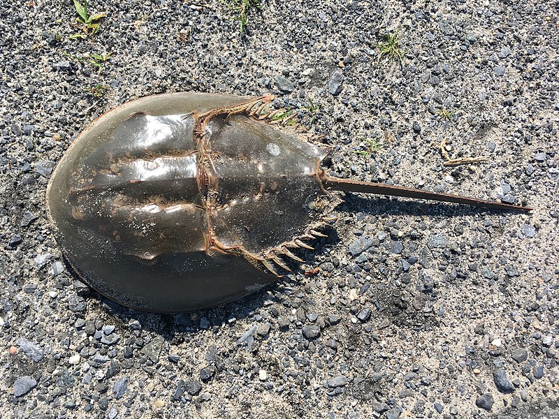
Paleozoic Paladin: Horseshoe Crabs and their Contribution to Vaccine Research

Hello, again, loyal MütterEDU readers. Editor Kevin here to introduce another piece from one of our amazing guest writers. Center for Education educator Joanna D’Avella prepared this article on the humble horseshoe crab and its huge contributions to vaccine research.

Does a prehistoric animal hold the key to worldwide vaccination? With its bright blue blood, horseshoe crabs (Limulus polyphemus) are the unsung hero of vaccine development in the 21st century as researchers tap into their evolutionary immune system to save the lives of millions. 

Atlantic Horseshoe Crab (Limulus polyphemus) (Image Source: Kaldari, via Wikimedia Commons, Public Domain)

How Does a Vaccine Get Approved?

According to the Centers for Disease Control and Prevention, only after rigorous amounts of testing throughout a variety of stages will a vaccine warrant FDA approval. These stages include the exploratory stage, pre-clinical stage, clinical development, regulatory review and approval, manufacturing, and quality control.

Some stages, similar to the development and manufacturing of the COVID-19 vaccine, can be done in conjunction with each other. This typically happens under an emergency authorization. The completion of stages in conjunction with one another does not reduce the efficacy of the vaccine due to the level of testing that is done prior to distribution.  

Why Do We Need to Test Vaccines on Animals? 

It is imperative that scientists create an environment that mimics that of a human body without placing humans in danger. As a result, scientists use animals as a safer alternative to human testing. Mice, monkeys, dogs, cats, and rabbits were all viable options for scientific research for hundreds of years, especially in early medical history when human dissection was illegal in most countries. Rabbits are the most well-known animal for animal testing, especially during the development of the polio and rabies vaccines. Researchers would use the rabbit pathogen test to confirm if pathogens were present in the vaccine. These animals gave scientists a safer alternative than using human subjects in the early stages of the vaccine development. 

Why Horseshoe Crabs?

Horseshoe crabs are an old species, having been around for over 300 million years. As one of the last connections to the prehistoric era, horseshoe crabs developed disease-prevention measures to maintain their population. This begins with their open circulatory system and their bright blue blood. In “The Role of Horseshoe Crabs in the Biomedical Industry and Recent Trends Impacting Species Sustainability,” the authors summarize the incredible process horseshoe crab blood undergoes when introduced to toxins. 

“When these granular amebocytes come into contact with an endotoxin or 1,3ß-D-glucan (present in the cell walls of Gram-negative bacteria and fungi, respectively), a cascade of defense molecules is released, triggering coagulation and neutralization of the pathogens. The resulting clot effectively immobilizes the threat and prevents an infection from progressing beyond the wound (Isakova and Armstrong, 2003).”

Krisfalusi-Gannon, J., Ali, W., Dellinger, K., Robertson, L., Brady, T. E., Goddard, M. K., Tinker-Kulberg, R., Kepley, C. L., & Dellinger, A. L. (2018). The role of horseshoe crabs in the biomedical industry and recent trends IMPACTING Species Sustainability. Frontiers in Marine Science, 5. https://doi.org/10.3389/fmars.2018.00185

Clotting helps prevent the toxins from spreading throughout the animal and making them extremely ill. Once researchers understood they could harness this evolutionary process, they have been using it ever since. 

During the 1970s, researchers began using horseshoe crab blood to test for endotoxins. Endotoxins are a toxin present in the outer membrane of cells that are released once the cell begins to decompose or is destroyed. Once released and introduced outside the cell, these toxins will then present symptoms of disease, making the organism very sick. In hopes of removing this potential threat, medical professionals devised tests to prevent these endotoxins from entering the general public through medical equipment or vaccines that had been contaminated. This was typically done using animals such as rabbits but at the cost of the test subject. Researchers monitored the rabbits for symptoms after injection; if none manifested, then medical equipment or a batch of vaccines were considered safe. However, if the rabbit–similar to a canary in a coal mine–died shortly after being exposed, it was deemed unsafe. Once researchers understood the potential of the horseshoe crab blood, they developed the limulus amebocyte lysate (LAL) which according to a 2010 article in the Journal of Applied Sciences, “in general were 3 to 300 times more sensitive than the United States Pharmacopeial (USP) rabbit pyrogen test method.” This was a scientific breakthrough for vaccine development. It was a far more effective test that did not necessarily mean the loss of animal life. However this is debatable because approximately 15% of horseshoe crabs that have underwent blood collection passed away as a result. This test effectively replaced the rabbits, and put these Paleozoic paladins on the frontline of medical developments. 

COVID-19 and Horseshoe Crabs

As vaccines are developed all over the world to try and to stop the spread of COVID-19, horseshoe crabs and their blood are being used more than ever due to the sensitivity needed for the LAL test. This species was already under ecological strains due the environmental changes, overfishing, and overuse in synthesis of the LAL test. It is true that researchers do release horseshoe crabs after their blood has been collected, however, the survival rate is not high enough to sustain the current population. There is hope, however, as scientists are developing an synthetic alternative to the horseshoe crab blood that will potentially yield similar results.

Sources

Akbar John, B., K.C.A Jalal, Y.B. Kamaruzzaman, and K. Zaleha. “Mechanism in the Clot Formation of Horseshoe Crab Blood during Bacterial Endotoxin Invasion.” Journal of Applied Sciences 10, Iss. 17 (2010). 

Cox, Elizabeth. “Why do we harvest horseshoe crab blood?” Ted-Ed (September 21, 2017).

Dhnesha, Neel. “How the Race for a COVID-19 Vaccine Jeopardizes East Coast Shorebirds.” Audubon. November 20, 2020. Accessed September 7, 2021.  

Esteves, P.J., Abrantes, J., Baldauf, HM. et al. The wide utility of rabbits as models of human diseases. Exp Mol Med 50, 1–10 (2018).

Krisfalusi-Gannon, J., Ali, W., Dellinger, K., Robertson, L., Brady, T. E., Goddard, M. K., Tinker-Kulberg, R., Kepley, C. L., & Dellinger, A. L. (2018). The role of horseshoe crabs in the biomedical industry and recent trends IMPACTING Species Sustainability. Frontiers in Marine Science, 5. 

Merriam-Webster. (n.d) Endotoxins. In Merriam-Webster.com dictionary. Retrieved August 17, 2021.

Pavid, Katie (3 December 2020, Updated: 15 January 2021) “Horseshoe crab blood: The miracle vaccine ingredient that’s saved millions of lives.” Natural History Muse.

Vaccination Trial Phases (2021) Center for Disease Control. https://www.cdc.gov/vaccines/basics/test-approve.html

Victorian Mental Health and Women, Part Three: Treating Depression

Hello, again, loyal readers, editor Kevin here welcoming you to guest writer Isabel DuBois’ third installment in a three-part series examining the history of mental health treatment for women in the 19th century. If you haven’t read parts one and two, I encourage you to check them out.

What we recognize today as depression was, in the Victorian era, popularly known as melancholia or melancholy. The name “melancholia” comes from the old medical belief in the four humours. Early medicine believed that every disease or ailment was caused by an imbalance in one or more of the four basic bodily liquids, or humours. According to Hippocrates and subsequent tradition, melancholy was caused by an excess of black bile, hence the name, which means “black bile” in Greek. A person whose constitution tended to have a preponderance of black bile had a melancholic disposition. Melancholy, like depression, ranged in seriousness from mild and temporary bouts of sadness to longer, more extreme episodes, characterized by insomnia, lack of appetite, and suicidal thoughts. Symptoms of melancholy were easy to recognize, although medical opinions often differed on what caused the condition. As a result, treatment plans for the melancholic patient varied. In an article in the 1850 edition of the People’s Medical Journal, Dr. Thomas Harrison Yeoman shared a catalogue of symptoms for melancholy, writing that “the leading characteristics of melancholy are—a love of solitude, gloom, fear, suspicion, and taciturnity.”

Albrecht Dürer, Melancholia I (1514), Engraving. Image Source: Metropolitan Museum of Art

Many medical practitioners of the time found it useful to divide melancholy into categories by symptom. This served to separate the more severe forms of melancholy, such as those accompanied by violent outbursts, mania, or delusions, from the more “ordinary” forms of melancholy in which the patient was merely reclusive and sad. In his 1871 book Insanity and its Treatment, Dr. G. Fielding Blandford classified melancholy as being either acute or subacute. Dr. Thomas Yeoman goes a step further in his book, Encyclopaedia of Domestic Economy, dividing melancholy into four separate types: 1) Gloomy Melancholy; 2) Restless Melancholy; 3) Mischievous Melancholy; and 4) Self-Complacent Melancholy.

Unfortunately for Victorian doctors and their patients, there were no antidepressant drugs available in the nineteenth century. Instead, doctors generally treated melancholy by recommending specialized diets and regimens of rest and relaxation via the rest cure, a treatment created by Silas Weir Mitchell, which consisted of weeks of forced feedings and bed rest with no mental stimulation. Some doctors would also recommend the “shower bath,” advising patients to shower often and, afterward, to “rub the whole body well with coarse flannel.” This was a much less traumatic option than the water therapy offered at some asylums, wherein patients were plunged into cold baths or, as in one case related by Dr. John Bell in his 1859 book A Treatise on Baths, “bound in a cart, stripped, and blindfolded” and then subjected to “a great Fall of Water” from twenty feet above. Some Victorian doctors went further with their treatments, advising their melancholy patients to drink alcohol, to take morphine, or even, in the case of women (if they were single) to get married and start a family. Dr. Blandford recommended a diet which featured alcohol at almost every meal, followed by a dose of chloral or morphia, intense sedatives, at night to help the melancholic patient sleep.

While great strides have been made in the field of psychology, we can’t dismiss entirely the Victorian efforts to understand the mind. Indeed, compared with the early asylums—rough, brutal places where the most disturbed patients were chained in windowless rooms with straw bedding—the mid-Victorian era was positively progressive. Theories that still hold today, such as the value of occupational therapy, were becoming fashionable. It was here that the shift away from the idea of control from without, via chains and shackles, and towards a patient’s control from within, via treatment or cure, began.

Sources:

Bell, John. A Treatise on Baths: Including Cold, Sea, Warm, Hot, Vapour, Gas, and Mud Baths. Philadelphia: Lindsay & Blakiston, 1859.

Blandford, G. Fielding. Insanity and its Treatment. Philadelphia: Henry C. Lea, 1871.

Flint, Austin. Clinical Medicine: A Systematic Treatise on the Diagnosis and Treatment of Diseases. Philadelphia: Henry C. Lea, 1879.

Matthews, Mimi. “A Cure for Melancholy: Victorian Medical Advice on Treating Depression.” MimiMatthews.com. April 3, 2017. Accessed August 20, 2021.

Maudsley, Henry. The Physiology and Pathology of the Mind. New York: D. Appleton and Company, 1872.

Nasim Jumana, Rumaisa. Mental Illness and Psychiatry in the Victorian Era: An Analysis of the Prevailing Power Dynamics Between Women and Male Authority Figures Through Gilman and Freud. City University of New York Masters Thesis. 2019.

Webster, Thomas. Encyclopaedia of Domestic Economy. New York: Harper & Brothers, 1855.

Yeoman, Thomas Harrison. The People’s Medical Journal, and Family Physician, Vol. L. London: George Vickers, Strand, 1850.

How We Spent Our Summer: The George and Judy Wohlreich Junior Fellows Program

It has been a very exciting summer for the Center for Education youth programs. On June 25, 2021, The College of Physicians of Philadelphia officially announced the creation of the George and Judy Wohlreich Junior Fellows program. Formerly the Karabots Junior Fellows and the College Junior Fellows program, the George and Judy Wohlreich Junior Fellows is a three-year summer and after-school program for Philadelphia high school students from historically excluded communities who are interested in careers in healthcare and medicine.

In August, we were happy to welcome the latest cohort of the program. A small group of students from all across the city came together to participate in an intensive two-week summer program. This year’s theme was “Lessons of 2020,” taking a closer look at the tumultuous year and a half we have all been experiencing through the lens of health. Some of the topics addressed included how diseases behave; lessons learned, and not learned, from past pandemics such as the 1918 influenza and 1980s AIDS pandemics; the social determinants of health, the impact of the COVID-19 pandemic on historically marginalized or excluded communities, such as incarcerated people and communities of color; and what it means to be a healthcare professional during a global pandemic.

A key element of the program is putting students in contact with healthcare professionals in the field. This summer, we were fortunate to have an exciting group of guest speakers who shared their professional expertise and life experiences with our students. These experts represented such fields as pathology, microbiology, immunology, public health, pediatrics, physical therapy, hospice and palliative care, internal medicine, and emergency medicine.

Our students also became experts in one aspect of the Mütter Museum. Working in small groups, they selected a specimen that is on display on Memento Mütter, our online exhibit, and prepared a mini tour on that specimen for guests from the Penn Summer Undergraduate Minority Resource (SUMR) program, a program of the Leonard Davis Institute of Health Economics at the University of Pennsylvania that introduces talented undergraduate students from underrepresented minority groups to research in health services, population health, and clinical epidemiology at Penn. Topics the students covered included the history of prosthetics, obstetrical equipment, various historic medications, and presidential assassin Charles J. Guiteau.

We are proud of our new students’ accomplishments and excited to offer them new educational opportunities during the school year.

If you are a Philadelphia high school student who will be starting their sophomore year during this coming school year, there are still a few spots available in our cohort. For more information on eligibility check out our website or our FAQ, or contact Kevin D. Impellizeri, PhD, Assistant Director of the Center for Education. You can also fill out an application HERE.