CEPI Curiosities: Shakespeare, Mozart, and the Hyrtl Skulls

Hello, fellow medico historians specializing in weirdness. Kevin again for another installment of CEPI Curiosities. I seem to have fallen into a rut of stories of mishandled corpses, as I have focused on an allegedly murdered President, a Presidential son/father son who became a medical specimen, and a fake Persian mummy. I had planned to go in a different direction this time around until I came across this recent story by The Guardian reporting that William Shakespeare himself may have been a victim of some cemeterial shenanigans.

Portrait of William Shakespeare

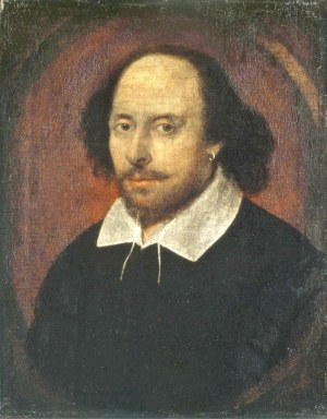
As part of a documentary on Shakespeare set to premiere in the UK this weekend, a team conducted an archaeological examination of William Shakespeare’s grave at the Holy Trinity Church at Stratford-Upon-Avon. Their analysis led them to the conclusion that the famous playwright’s skull was no longer with the rest of his remains. This confirmed a long-standing rumor dating back to the 19th century that Shakespeare’s skull had been taken by graverobbers in 1794.

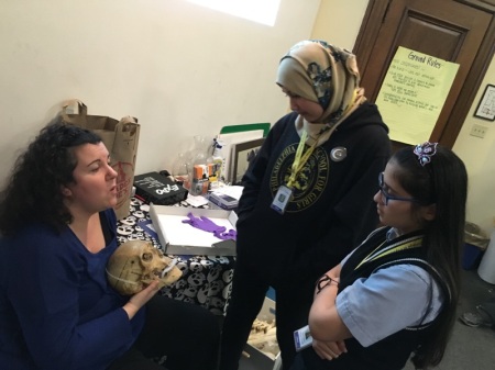
This shocking revelation of the possibly-headless Bard, whose grave site famously curses anyone for moving his bones, reminded me of a similar story of a famous artist whose skull turned missing that involved one of the Mütter Museum’s most famous scientists: Joseph Hyrtl. Hyrtl was a Viennese anatomist who made it his life’s work to disprove phrenology, the belief that the physical dimensions of the skull correlated with personality. To that end, Hyrtl collected and measured hundreds of European skulls. He concluded that the vast disparity in dimensions of European skulls disproved any common connections to personality. His work also disproved the inherent intellectual superiority of Caucasians over other races. Phrenology was commonly used as evidence to reinforce scientific racism, as evidenced by famous phrenologists such as Samuel George Morton of the Academy of Natural Sciences (whose own collection of skulls can be viewed on display at the nearby Penn Museum). In 1874, Hyrtl sold his collection of 139 skulls to the Museum, many of which you can view on display today.

Image of the Hyrtl Skull Collection at the Mütter Museum

Among the skulls that came into Hyrtl’s possession, rumor has it that one curious cranium belonged to none other than Wolfgang Amadeus Mozart. The legendary Austrian composer and musician died in poverty at the age of 35 and was buried in an unmarked grave in a Viennese common cemetery in 1791. Bodysnatchers allegedly removed his skull, which eventually came into the possession of Joseph’s brother Jacob, who willed it to his brother following his death in 1868.

However, before you rush to the Mütter Museum with your camera and your powdered wig, the alleged Mozart skull was not part of the collection Hyrtl sold to the College of Physicians. Rather, Hyrtl willed the skull to the Austrian city of Salzburg in the early 1890s. Following his death in 1894, the skull reportedly changed hands and eventually appeared in the collection of the International Mozarteum Foundation in Salzburg (read a detailed account here and here), where it remains to this day (although it is not on display to the public).

Black & white portrait of Mozart

Whether the skull really belonged to Mozart has never been definitively proven. The Mozarteum skull has been subjected to numerous forensic tests over the years, including DNA analysis and two separate facial reconstructions. These tests revealed differing conclusions over its authenticity. There is also some scholarly debate over whether the skull at the Mozartium is the one previously owned by Hyrtl, as there are inconsistencies between Hyrtl’s descriptions and the Mozarteum skull. We may never know whether the skull really was Mozart’s.

In the meantime, be sure to not lose your head and come to the Mütter Museum to check out the Hyrtl Skull Collection and many other specimens from the history of medicine. Until next time!


A Symposium for Providers: Understanding Gender Identity & Development for Transgender Youth in 2016

Logos of the sponsors for the March 17 Gender Symposium

Tomorrow the College of Physicians of Philadelphia will be hosting a symposium on gender development.

Nadia Dowshen, MD, and Linda Hawkins, PhD, MSEd, LPC, will lead a discussion on gender identity development and discuss how to make spaces and practices safer for trans, gender-variant, and gender-nonconforming (GNC) youth. Dr. Dowshen and Dr. Hawkins are the co-directors of the Gender and Sexuality Development Clinic at the Children’s Hospital of Philadelphia (CHOP). The event will take place in Mitchell Hall at the College of Physicians of Philadelphia and is scheduled to start at 6:30 PM.

After the presentations by the clinical staff, interns from the Mütter Museum’s Out4STEM Program will lead a session on the experiences of trans, gender-variant, and GNC youth, to highlight the need for competent care.

The event is open to the public (Tickets are FREE). Healthcare providers, educators, and youth-serving professionals are especially welcome.

The Karabots Fellows Become Bone Detectives

Members of the Karabots Junior Fellows Program investigate the race, sex, and age of bones

Ever wonder how scientists are able to identify human bones? The students of the Karabots Junior Fellows Program need not wonder anymore. As part of their year-long study of forensic science, the Fellows recently learned about forensic anthropology (the science of identifying human skeletal remains) with resident expert Anna Dhody. Anna, the Curator for the Mütter Museum and a trained forensic anthropologist (astute followers of everything involving the Mütter Museum may recognize her from her regular appearances on the web series: Guess What’s on the Curator’s Desk), took the Fellows through the science of identifying human remains while telling stories of her experience as an expert in criminal cases involving human (or what appear at first glance to be human) bones.

Two of the Karabots Junior Fellows talk with Anna Dhody, Curator of the Mütter Museum, about forensic anthropology

Following their meeting with Anna, the students held a session with class regular and Mütter Museum Educator Marcy Engleman. Marcy demonstrated how to identify the race, sex, and age of human bones, encouraging them to draw their own conclusions based on observations made on replicas of the real thing.

Mütter Museum Educator MArcy Engleman explains how to identify skeletal remains

If you are a teacher and interested in your students taking part in the Bone Detectives lesson, you can schedule a session as part of a field trip to the Museum. Happy bone detecting!

Philly Youth Navigate the Trenches at Fort Mifflin

Our students learn about the Battle of Verdun from a french re-enactor at Fort Mifflin

On March 5, students from the Teva Pharmaceuticals Internship and Karabots Junior Fellows Program took a field trip out of Philadelphia proper to visit historic Fort Mifflin. Fort Mifflin was commissioned in 1771 as a coastal defense Fort to protect Philadelphia from invaders and pirates. During its 183 years as an active military base, it was the site of the largest bombardment of the Revolutionary War (November 10-15, 1777), rebuilt as part of America’s first coastal defense system (the First and Second American Systems), served as a Union Army prison during the Civil War, and acted as an ammunition depot for the United States Navy during the First and Second World Wars. Today it serves as a historic site and museum.

A Teva fellow learns about the life of a World War I soldier from a German re-enactor at Fort Mifflin

The students visited the Fort to attend an event commemorating the 100th anniversary of the Battle of Verdun (Feb-Dec 1916), a crucial battle during World War I. Students got the chance to interact with reenactors from both the Allies and Central Powers to learn about the history of the Battle, the equipment soldiers carried and the human toll of the Battle, which left roughly 300,000 dead and 750,000 wounded. While at the Fort, they also explored the site, which includes underground shelters, historic ammunition magazines, and a long-buried Civil War prison cell that once held a Union prisoner who was later executed at the Fort. They even got to witness the firing of a Revolutionary War cannon! Braving cold and muddy conditions (they quickly learned why Fort Mifflin earned the nickname “The Mud Island”), everyone expressed excitement over the trip.  After an exciting visit, they returned to the College with new-found perspective on “The Great War.”

Cannon firing demonstration at Fort Mifflin

CEPI Curiosities: The Washing Away of Wrongs

Kevin here again, and I hope your hunger for medico-historico oddities has yet to be satiated because I am back for another installment of CEPI Curiosities. Last time, we investigated the case of the Persian Princess, a fake historic artifact that led to a real murder investigation. This offers a great jumping point to bring us back to the subject of study for the current cohort of the Karabots Junior Fellows Program: forensic science.

The twelfth and thirteenth centuries were an instrumental period in the history of forensic science, as several cultures lent more scientific weight to death investigation. In 1194, England established an official office of the coroner to investigate cases of sudden death or murder (As an aside early English coroners also had the power to perform arrests as well as the power to collect taxes). Around the same time in Bolonga (in modern-day Italy), the first criminal cases emerged where medical professionals were brought in as expert witnesses. Medical autopsies gradually gained favor in Europe around the same time as scientists challenged earlier taboos against examining human remains.

However, the first formalized procedures for forensic investigation originated in Song Dynasty China.  In 1247 Song Ci (Sung Tzu in Wade-Giles), wrote and published the first known forensic handbook: Collected Cases of Injustices Rectified, or The Washing Away of Wrongs (Xi Yuan Ji Lu). Directed at local coroners, the book mapped out detailed guidelines for investigating sudden or mysterious deaths. At a time when coroners required no formalized training, his guidelines provided clear methods for conducting investigations.

Song based his guidelines on practical observation and inferences based on crime scene evidence and body examination. He explained how to examine bodies under certain conditions, such as buried vs. non burial, various stages of decomposition, and skeletal remains. He provided detailed guidelines for conducting autopsies, including diagrams. He showed how to identify evidence of stabbings, blunt force trauma, strangulation, the presence of various poisons, and even lightning strikes, tiger bites, and death from excessive eating, drinking, or sex. He also went into extensive detail on how to determine murder from suicide. Song Ci’s treatise served as the standard manual of forensic investigation in China for centuries and was translated into numerous languages, including English, German, French, and Japanese.

Nomenclature of Human Bones from the 1843 edition, Image in the Public Domain

Nomenclature of Human Bones from the 1843 edition, Image in the Public Domain

As interesting as The Washing Away of Wrongs is on its own as a practical manual of crime scene investigation, this wouldn’t be CEPI Curiosities without some medical weirdness. In addition to all the benefits Song Ci’s work gave for the novice or experienced coroner, Song also gave advice on how to undo the effects of death. In his penultimate section, titled “Methods for Restoring Life,” Song enumerated various tried and true methods to reanimate dead subjects. These remedies covered different specific instances of death, such as drowning, freezing, and death from fright and often involved massaging the limbs, blowing air into the victim’s ears, or forcing the (dead) subject to ingest something. For example, a victim of what Song described as the “five deaths” (death from childbirth, fright, strangulation, nightmare, and drowning, presumably not all at the same time) could be revived, even if they had been dead for more than a day, provided the following:

“First take the dead person and place him on the ground with knees bent, like the posture of a Buddhist monk in meditation. Have someone take the dead person’s hair and pull it down towards the ground. Using a bamboo or paper tube, blow a quantity of raw powdered Pinellia ternata [an herb which is a deadly toxin in its raw form]…into the nose. If he revives, administer a dose of raw ginger juice, which will counteract the poisonous effects of the Pinellia” (Sung Tz’u, The Washing Away of Wrongs, Brian E. McKnight, trans., Ann Arbor: Center for Chinese Studies, the University of Michigan, 1981, 161).

Among my favorites is a remedy he prescribed for sudden deaths by “Repulsive Factors” (most likely a heart attack) which involved “thrust[ing] the yellow heart of a leek six or seven inches into the nostril (the left for men, the right for women), causing the blood from [the part of the nostrils] between the eyes to flow. The victim will then revive”(Ibid. 158-159).

If you would like to know more, the Library of the College of Physicians has a copy of the English translation. It provides a fairly quick and definitely informative and entertaining read. Until next time!

The Teva and Karabots Fellows Explore the Small World of Microphotography

Teva and Karabots students explore the Small Worlds Exhibit at the Wistar Institute

Recently, students in the Karabots and Teva programs took a trip to the Wistar Institute where they got to get a close look at a mixture of photographic art and microscopic science.

The Wistar Institute was founded in 1892 as a anatomical museum (not unlike the Mütter Museum) and evolved into a state-of-the-art biomedical research facility. Today the site is designated as a National Cancer Institute Cancer Center in basic research. Scientists at the Wistar have played a role in identifying the genes associated with a variety of cancers as well as breakthrough research into vaccines, antibodies and other aspects of medicine.

The Wistar is also a host for the Nikon Small World Exhibition. The Small World Exhibit is the culmination of an annual photography contest where contestants submit artistically-treated microscopic images. The images cover a variety of subject matter, including plants, animals, insects, and microorganisms (see the 2015 winners). Through complex photo editing techniques, the photographers bring the microscopic images to life in vibrant color.

James Hayden presents different micro photographic techniques to the Karabots and Teva fellows

The students met with James Hayden, Managing Director of Imaging Shared Resource, who took them through the exhibition and the technical and scientific work that made it possible.

Teva and Karabots students explore the Small Worlds Exhibit at the Wistar Institute

If you would like to explore the Small World exhibition for yourself, it will be at the Wistar Institute through March 6 before heading to the Texas Museum of Science & Technology in Cedar Park, TX (full schedule). For more events and activities at the Wistar Institute, check their events schedule.